
A 13.5-year-old spayed female Beagle presented to Ruby Veterinary Urgent Care for evaluation of acute left forelimb lameness or a sudden limp on her front leg.
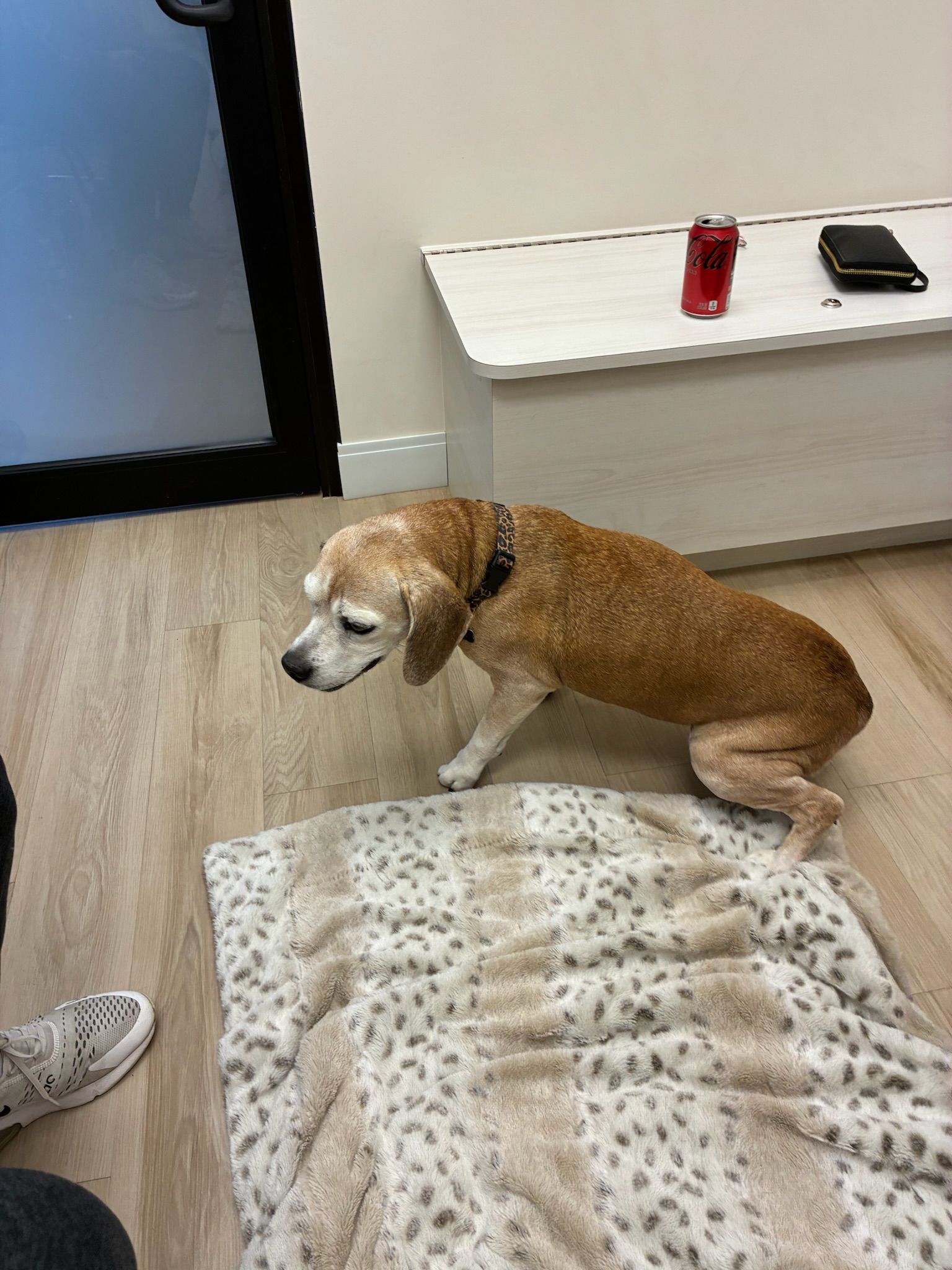
According to her owners, the lameness came on abruptly, with no known history of trauma such as a fall or accident. While the dog had previously experienced intermittent rear limb lameness believed to be related to arthritis (age-related joint wear and tear), this episode involved a new and more severe limp affecting the front limb, which raised additional concern.
Aside from the limp, the dog was otherwise doing well at home. She was reported to be bright, alert, and responsive, with normal appetite, water intake, and energy level.
Physical Examination Findings
On examination, the patient remained bright, alert, and responsive, indicating that she was feeling well overall. Her vital signs, such as heart rate, respiratory rate, and temperature, were all within normal limits.
During the orthopedic exam (an assessment focused on bones, joints, and soft tissues), several important findings were noted:
- Moderate swelling of the left antebrachium (The antebrachium is the portion of the front leg between the elbow and the wrist.)
- Pain on palpation, meaning the dog reacted when the area was gently pressed
- A superficial skin abrasion, or small scrape, over the swollen region
- Palpable crepitus, which feels like crackling or bubble-wrap under the skin and is characteristic of air trapped beneath the skin.
No gross instability (looseness of the bones or joints), angular deformity (abnormal bending), or neurologic deficits were detected. The remainder of the physical examination was unremarkable.
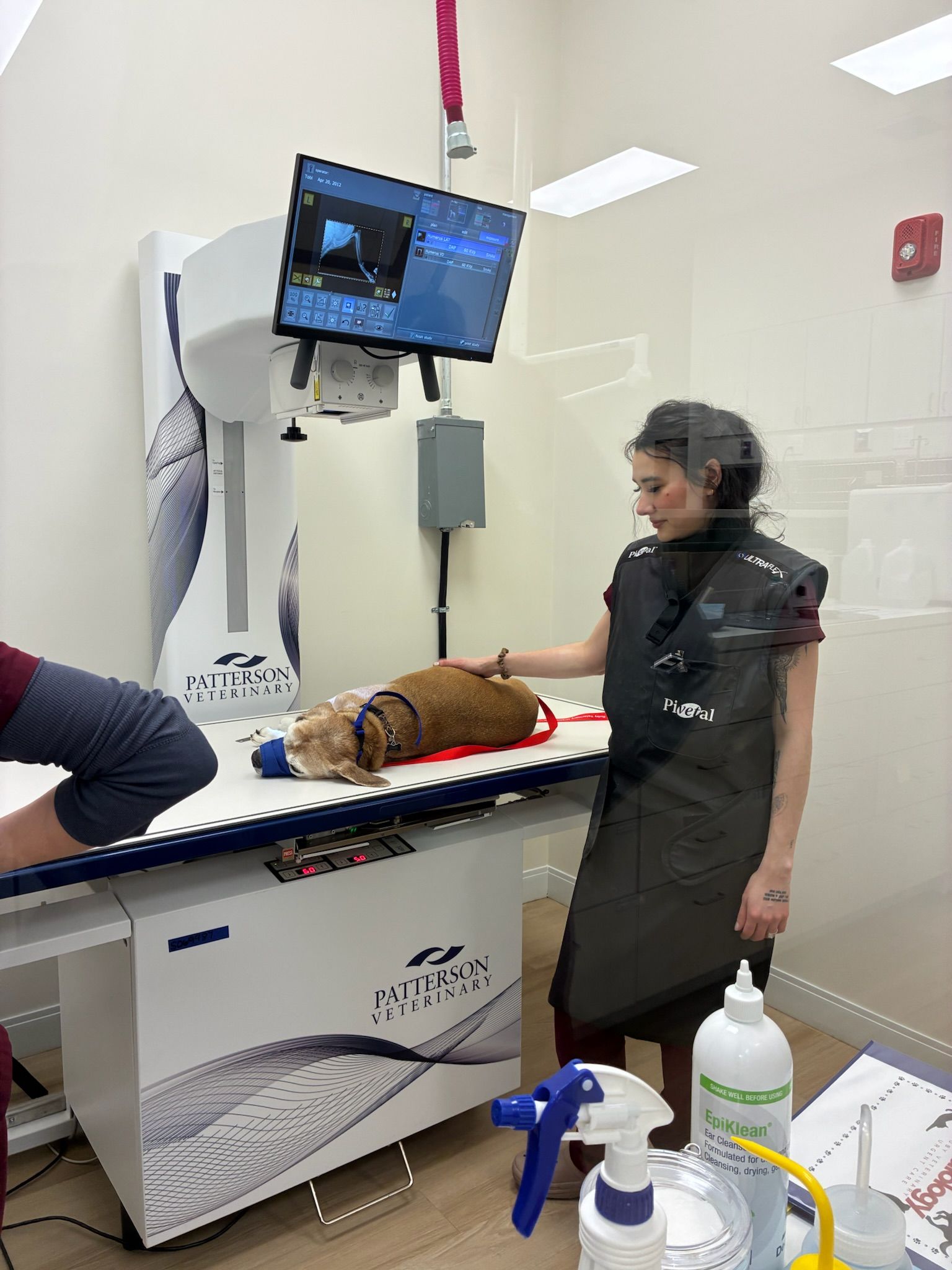
Diagnostic Imaging Indicated No Fractures
Radiographs (X-rays) of the left forelimb were obtained to further evaluate the cause of the swelling and lameness.
Imaging revealed:
- Diffuse soft tissue swelling, meaning inflammation of the muscles and tissues rather than the bones
- Subcutaneous emphysema, which refers to air trapped within the tissues just under the skin
Importantly, no fractures, joint dislocations (luxations), or abnormalities of the underlying bones were identified.
Diagnosis
Based on the physical examination and radiographic findings, the patient was diagnosed with:
Soft tissue trauma of the left antebrachium with associated subcutaneous emphysema
In plain terms, this means the dog likely sustained an injury to the soft tissues of her front leg, such as muscles or connective tissue, with air becoming trapped under the skin, likely through a small wound or trauma.
Treatment Plan and Outcome
Because the injury did not involve broken bones or systemic illness, the patient was treated conservatively on an outpatient basis, meaning hospitalization was not required.
The treatment plan included:
- Analgesic medications to control pain and keep the dog comfortable
- Oral antibiotics to reduce the risk of infection, particularly given the presence of a skin abrasion and trapped air
- Strict rest and close monitoring at home to allow the tissues to heal and to watch for any signs of worsening
At the time of discharge, the patient remained stable, comfortable, and clinically well.
Discussion: Understanding Subcutaneous Emphysema in Dogs and Cats
What Is Subcutaneous Emphysema?
Subcutaneous emphysema is the presence of air within the tissues beneath the skin. When touched, it often feels like crackling or popping under the fingers.
In dogs, this can occur for several reasons, including:
- Minor penetrating wounds or trauma that allow air to enter beneath the skin
- Air tracking from nearby structures after an injury
- Infection with gas-producing bacteria, which create gas as part of their metabolism
Certain bacteria such as Clostridium species, Escherichia coli, and Streptococcus species can produce gas in low-oxygen environments. When this happens, gas can accumulate in the tissues, leading to swelling, discomfort, and crepitus.
When Is Trapped Air a Medical Emergency?
Although uncommon, gas-producing infections such as clostridial myonecrosis (often referred to as gas gangrene) are medical emergencies. These infections progress rapidly and can cause severe tissue destruction and systemic illness.
Red flags for serious infection include:
- Rapidly worsening pain and swelling
- Skin that feels hot or appears discolored
- Lethargy, fever, or other signs of systemic illness
- Gas seen on X-rays extending into deeper tissues, such as muscle
In this case, the gas was limited to the subcutaneous tissues (just under the skin), with no muscle involvement, no bone abnormalities, and no signs of systemic illness. The dog remained bright and comfortable, making a severe gas-producing infection less likely at the time of evaluation.
However, the presence of a skin abrasion suggested a possible entry point for bacteria or air. For that reason, early antibiotic therapy and close follow-up were recommended to reduce the risk of progression.
Clinical Takeaway for Pet Owners
Lameness in dogs is often caused by relatively minor issues such as soft tissue strain or arthritis. However, as this case demonstrates, sudden or severe limping, especially when accompanied by swelling or pain, deserves prompt veterinary evaluation.
Diagnostic tools such as physical examination and X-rays allow veterinarians to distinguish between:
- Minor, self-limiting injuries
- Soft tissue trauma
- More serious infectious or emergent conditions
Early evaluation helps ensure that potentially serious problems are identified quickly and treated appropriately.
With extensive emergency and urgent care training, the team at Ruby Veterinary Urgent Care is equipped to assess pets with both obvious injuries and subtle changes, helping families make informed decisions when care cannot wait.