
Imagine this: you go to bed, but in the middle of the night you're woken up by the sound of constant head shaking and scratching. You think, “It’s probably an ear infection. I’ll take him to the vet in the morning.” But when you wake up, you can’t believe your eyes. Your dog’s ear flap is swollen and feels like a pillow. What happened?
While not all ear infections lead to aural hematomas, they are a common complication of chronic head shaking and scratching. In this article, we’ll explain what an aural hematoma is, how it develops, and what treatment options are available to help your pet heal.
What Is an Aural Hematoma?
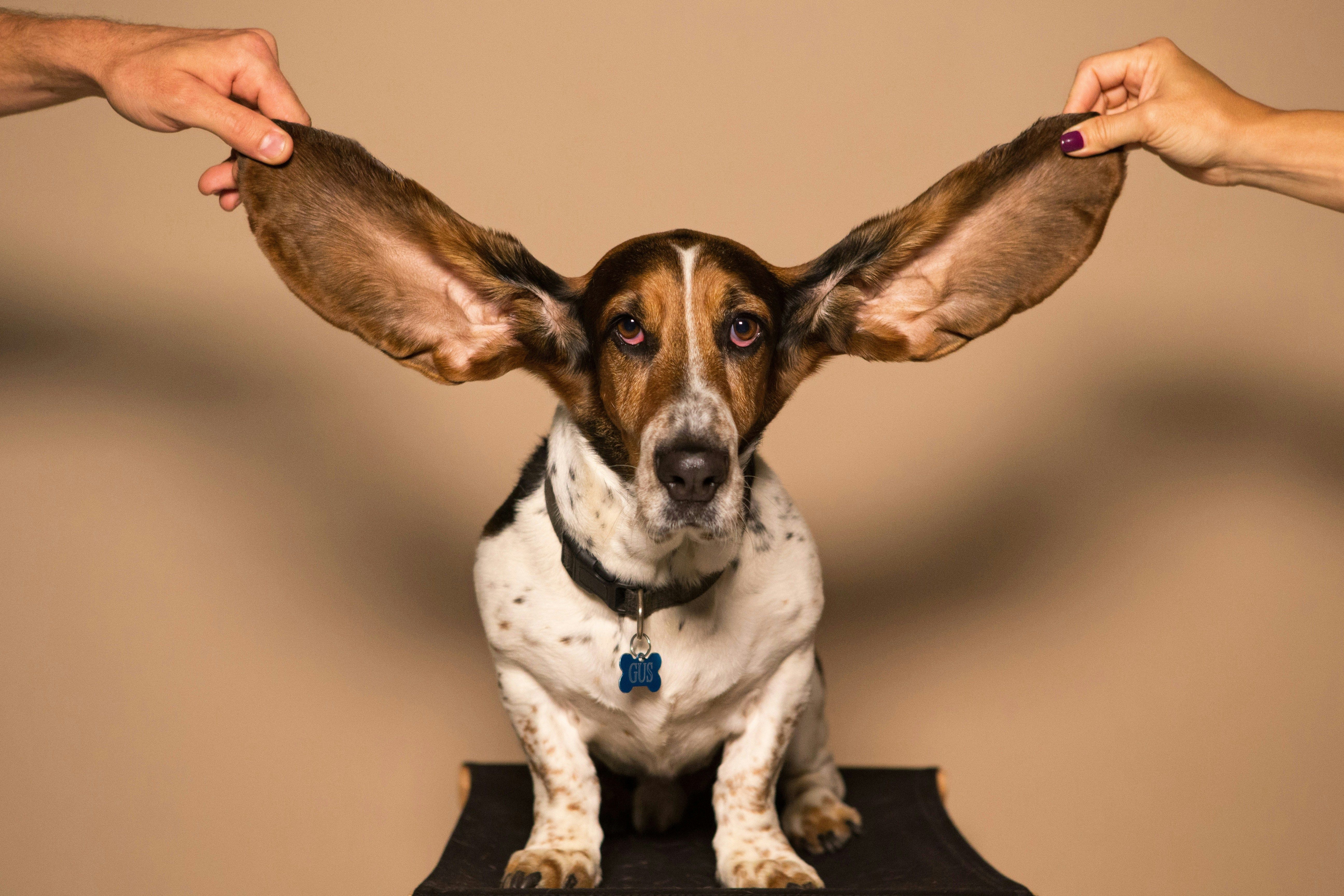
An aural hematoma is a pocket of blood that collects between the skin and cartilage of the ear flap (also called the pinna). This condition is most common in dogs but can also occur in cats. It often causes swelling, tenderness, and a 'ballooned' or 'puffy' ear appearance.
What Causes Aural Hematomas in Dogs and Cats?
Aural hematomas are usually caused by trauma to the ear, most often from repeated head shaking or scratching. When this happens, the small blood vessels in the ear flap rupture, allowing blood to leak into the space between layers of cartilage and skin.
The underlying trigger is often something that makes the ear itchy or uncomfortable, such as:
- Ear infections
- Allergies
- Ear mites
- Foreign bodies (like grass seeds)
- Dermatitis or skin irritation
In some cases, rough play or physical trauma to the ear can also lead to hematoma formation.
What Does an Aural Hematoma Look Like?
Aural hematomas typically appear as sudden swelling on one or both ears. The ear flap may feel soft and squishy or firm, depending on how much blood has accumulated. You might also notice:
- Redness and warmth at the site
- Fluid-filled appearance
- Discomfort or flinching when the ear is touched
- Head shaking or tilting
- Ear discharge if an infection is present
If left untreated, the body may eventually reabsorb the blood on its own. However, this often results in a crinkled or wrinkled appearance known as 'cauliflower ear.' While this is usually cosmetic, it can become permanent.
How Aural Hematomas in Pets are Diagnosed
A veterinarian will diagnose an aural hematoma through a physical examination. However, the priority is determining why it developed in the first place. Your vet will examine the ear canal with an otoscope to look for signs of infection, debris, or parasites. If ear discharge is present, a sample may be analyzed under a microscope to check for yeast, bacteria, or ear mites.
Treatment Options for Aural Hematomas
The best treatment approach involves addressing both the hematoma itself and the underlying cause. This reduces the risk of recurrence and helps your pet heal faster.
1. Surgical Drainage and Repair
Surgery is the most effective and long-lasting solution, especially for larger hematomas. The vet makes a small incision to drain the blood and clots, then places multiple sutures in a quilted pattern to prevent fluid from re-accumulating. This procedure requires general anesthesia and may not be suitable for pets with certain health risks. However, it offers the best chance for full recovery with minimal cosmetic changes.
2. Cannulation
Cannulation involves placing a small tube into the hematoma to allow continuous drainage over time. This technique is less invasive than surgery but may require close monitoring and follow-up care.
3. Needle Aspiration
This method involves using a syringe to draw out the fluid. While it's quick and relatively easy, aspiration is often a temporary solution. To be effective, the ear must be wrapped snugly against the head to prevent the hematoma from refilling. The ear canal is left exposed to allow for treatment of any infection or infestation.
4. Medical Therapy
Injections of corticosteroids can reduce inflammation and promote healing. This treatment is sometimes used in combination with drainage procedures or on its own for smaller hematomas. Cold laser therapy or photobiomodulation (such as Phovia) may also be used to reduce swelling, promote tissue repair, and relieve pain.
5. Benign Neglect (Watchful Waiting)
In select cases, small hematomas may resolve on their own. This conservative approach is only considered if the pet is not in pain and the hematoma is not interfering with the ear canal. However, this option may result in a permanent deformity (cauliflower ear) and can take weeks or months to heal. If the hematoma is large or painful, treatment is usually recommended to avoid complications like ear canal obstruction or chronic discomfort.
Prognosis and Prevention
With prompt veterinary care, the prognosis for aural hematomas is excellent. Once the hematoma is treated and the underlying cause is addressed, most pets recover well. To help prevent aural hematomas:
- Treat ear infections and allergies promptly
- Use parasite prevention to protect against mites
- Keep your pet’s ears clean and dry
- Monitor for excessive scratching or head shaking
- Avoid rough play that could injure the ears
Final Thoughts
An aural hematoma may look dramatic, but it is a treatable condition with a range of medical and surgical options available. The key is to act quickly. If your pet develops a swollen, painful ear or shows signs of discomfort, have them evaluated by a veterinarian right away. At Ruby Veterinary Urgent Care, we’re here seven days a week to help with urgent concerns like aural hematomas, ear infections, or anything else causing your pet pain or distress. Our experienced team can guide you through the best treatment options and help your pet feel better, faster.